
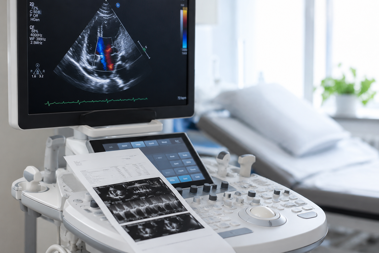
An echocardiogram, often called an “echo,” is a noninvasive imaging test that uses high-frequency sound waves to create real-time pictures of your heart. Unlike X-rays or CT scans, an echocardiogram does not use radiation.
This test allows your provider to see how your heart is functioning, evaluate blood flow, and identify abnormalities that may be causing symptoms or increasing your risk for cardiovascular disease.
An echocardiogram is commonly performed in the office and is one of the most valuable diagnostic tools available in cardiology.
our provider may recommend an echocardiogram if you have symptoms that suggest a heart condition or if another test indicates that additional evaluation is needed. It is also frequently used to monitor patients with known cardiovascular disease and evaluate how well treatments are working over time.
Your provider may also order an echocardiogram before certain surgeries or procedures to better understand your heart function and determine whether any additional precautions are needed.
An echocardiogram provides detailed information about many aspects of heart health that cannot be determined through a physical exam alone. The images help your provider evaluate both the structure of your heart and how effectively it is pumping blood throughout your body.
A standard transthoracic echocardiogram is performed in a comfortable outpatient setting and usually takes between 30 and 60 minutes. The exam is painless, requires no incisions, and most patients can resume normal activities immediately afterward.
Preparation – You’ll change into a gown, and small adhesive electrodes may be placed on your chest to monitor your heart rhythm throughout the examination.
Image Collection – A cardiac sonographer will apply a warm gel to your chest and gently move a handheld ultrasound transducer across several areas. The sound waves create detailed images of your heart that appear instantly on a monitor.
During the exam, you may be asked to briefly hold your breath or change positions to allow additional views of the heart.
After the Test – Your provider may ask you to briefly change positions or hold your breath so additional images can be captured from different angles.
Once the exam is complete, you can immediately return to your normal daily activities.
Different types of echocardiograms may be recommended depending on your symptoms and medical history.
Transthoracic Echocardiogram (TTE) – The most common type of echocardiogram, performed by placing an ultrasound probe on the chest.
Stress Echocardiogram – Images are taken before and after exercise or medication that increases your heart rate to evaluate how your heart performs under stress.
Transesophageal Echocardiogram (TEE) – A specialized ultrasound probe is passed into the esophagus to obtain highly detailed images of the heart when additional visualization is needed.
Your provider will recommend the test that best fits your condition.
After your test, your cardiovascular specialist carefully reviews the images and measurements.
Your physician will explain the findings, discuss whether additional testing is needed, and recommend the next steps for your care.
An echocardiogram is one of the safest and most informative diagnostic tools available in cardiovascular medicine. It provides detailed information while remaining comfortable and convenient for patients.
Noninvasive and Comfortable – No surgery, needles, or incisions are required for a standard echocardiogram, making it a low-risk diagnostic procedure.
Radiation-Free Imaging – Unlike CT scans or certain other imaging tests, echocardiography uses ultrasound rather than radiation, making it safe for repeated evaluations when needed.
Real-Time Heart Function – Providers can watch your heart beating in real time, allowing them to evaluate pumping strength, valve movement, and blood flow during the examination.
Early Detection of Heart Disease – Many heart conditions can be identified before symptoms become severe, allowing treatment to begin earlier and potentially improving long-term outcomes.
Personalized Treatment Planning – The information gathered during an echocardiogram helps your cardiovascular specialist make informed decisions about medications, additional testing, lifestyle recommendations, or interventional procedures.